Hematology and anemia in animals: what you need to know
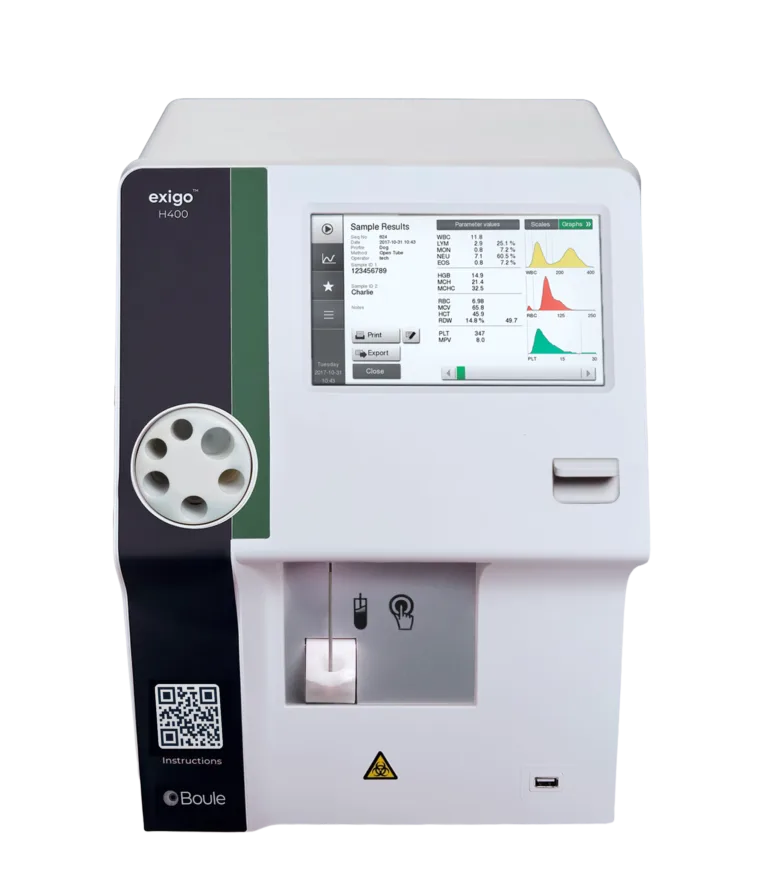
In this article, we cover the key indicators to consider when screening for and investigating anemia in animals. We also review how automated hematology solutions, such as the Exigo H400, can provide relevant clinical parameters to support efficient and cost-effective anemia screening.
Anemia is a common diagnosis in animals and humans alike. It can range from chronic anemia, which may cause weakness or lethargy, to acute anemia, which can result in shock and can be life-threatening. Identifying the underlying cause and determining whether anemia is regenerative or non-regenerative are essential steps in patient management.
Identifying the cause of anemia typically requires a combination of anamnesis (patient history), physical examination, and clinical testing such as hematology. While microscopy is required to assess specific red blood cell (RBC) morphologies, automated hematology analyzers are widely used as initial screening tools that provide valuable quantitative information. Hematology screening is therefore often the first step in anemia investigations, offering an overview of hematological status and absolute values of key cell populations.

Anemia screening in animals: key hematological indicators
Anemia is defined as a reduced oxygen-carrying capacity of red blood cells. RBC indices are therefore central to diagnosis. Parameters of particular relevance include hematocrit (HCT), mean cell volume (MCV), mean corpuscular hemoglobin (MCH/MCHC), and red blood cell distribution width (RDW).
Hematocrit (HCT)
HCT represents the volume percentage of RBCs in circulation. While reference ranges vary with species, breed, age, and physiological status, a reduced HCT is a strong indicator of anemia and can also provide information about severity.
Mean corpuscular volume (MCV)
MCV reflects the average size of RBCs. Immature RBCs, such as reticulocytes, are typically larger than mature RBCs, and their presence increases MCV. Other clinically relevant findings include microcytosis associated with iron deficiency (low MCV) and macrocytosis associated with folate or vitamin B12 deficiency (high MCV).
Mean cell hemoglobin (MCH) and mean cell hemoglobin concentration (MCHC)
MCH reflects the average hemoglobin content per RBC and often follows changes in MCV. However, MCH may be affected by analytical interference, such as lipemia or intravascular hemolysis, which can lead to falsely elevated hemoglobin values. Comparison with MCV, which is unaffected by these factors, is therefore important.
An increased MCHC is always an artifact and may result from Heinz bodies, RBC agglutination, or excess EDTA. A decreased MCHC may be observed in regenerative anemia or iron-deficiency anemia.
Red blood cell distribution width (RDW)
RDW reflects variation in RBC size and, together with the RBC histogram, provides insight into the presence of reticulocytes. Increased RDW and a broader histogram may indicate anisocytosis. A left-shifted histogram with increased RDW may suggest microcytosis, such as that observed in iron deficiency, where a larger proportion of small RBCs are present.

Screening made simple
In most clinical settings, 3-part, 4-part, and 5-part hematology analyzers provide sufficient information for initial anemia screening. Regardless of analyzer type, microscopy remains the next diagnostic step if abnormal values are detected. The key requirement for screening is therefore the analyzer’s ability to detect and flag abnormal samples.
In this context, the Exigo H400 can be used as a general screening tool for anemia investigations. Compared with more complex laser-based flow cytometry systems, impedance-based 3- and 4-part analyzers are often easier to maintain. The Exigo H400’s 4-part analysis includes an eosinophil count while retaining the robustness and lower maintenance requirements typical of impedance-based systems.
For additional information on anemia screening and hematology analysis, further resources are available upon request.